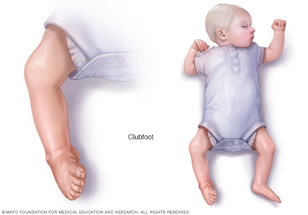
But, she did say that the ultrasound tech noticed baby girl has one club foot I know this can be common & Introduction The second trimester ultrasound is commonly performed between 18 and 22 weeks gestation Historically the second trimester ultrasound was often the only routine scan offered in a pregnancy and so was expected to provide information about gestational age (correcting menstrual dates if necessary), fetal number and type of multiple pregnancy, placentalClubfoot is a deformity in which an infant's foot is turned inward, often so severely that the bottom of the foot faces sideways or even upward Most cases of clubfoot can be successfully treated with nonsurgical methods that include stretching, casting, and bracing
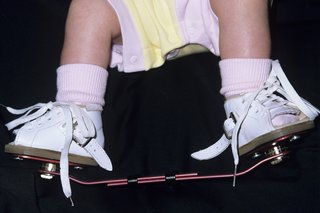
Club Foot Nhs
Baby ultrasound club foot
Baby ultrasound club foot- Babies who are born with a foot that's twisted inward and downward have a birth defect called clubfoot Find out what may cause it and how doctors fix it before babiesClubfoot is a birth defect of the foot that may affect your baby's ability to walk normally Clubfoot causes one or both feet to twist into an abnormal position, and can be mild or serious Learn how clubfoot is treated
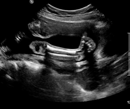


Congenital Talipes Equinovarus Radiology Reference Article Radiopaedia Org
An ultrasound at this gestational age will help confirm your due date, and it is part of the early risk assessment testing (ERA) for a chromosomal abnormality, such as Down syndrome The ultrasound technician will measure the fold on the back of the fetal neck Both a boy and a girl fetus have this fluidfilled space BACKGROUND AND PURPOSE Common prenatal ultrasound finding with varying severity and may be isolated or complex (presence of additional anatomical findings) There is controversy as to whether isolated clubfoot requires additional invasive testing to determine karyotype Weiner et al (Prenatal Diagnosis, 17) examined the diagnostic accuracy, related Why are baby's legs crossed during ultrasound?
Ultrasound introduces energy into the body This can heat body tissue, and can also produce small pockets of gas in body tissue or fluids (cavitation) It is not known what the longterm consequences of these effects might be for women and their unborn babies Human studies have not yet shown a direct association between pregnancy ultrasound The Clubfoot Chronicles Our clubfoot journey began on , when I was 19w3d pregnant with our fifth child I went to my OB/GYN's office for my routine week anatomy ultrasound, along with my husband and my two oldest children (We did not find out his/her gender, and refer to the baby as "Tiebreaker," since s/he has two brothers and The diagnosis was changed after followup ultrasound scan in 13 fetuses (25%), and the final ultrasound diagnosis was normal in one fetus, isolated club foot in 31 fetuses, and complex club foot in fetuses At birth, club foot was found in 79 feet in 43 infants for a positive predictive value of %
Club foot affects about 1 baby in every 1,000 born in the UK Both feet are affected in about half of these babies It's more common in boys Diagnosing club foot Club foot is usually diagnosed after a baby is born, although it may be spotted during the routine ultrasound I am 22 weeks pregnant with baby #2 (I have a 14 month old little girl no complications) At my week ultrasound, they found a Single Umbilical Artery and a problem with baby's kidney I went to get a high risk ultrasound done and they found that baby has a hole in her heart, a blockage in her kidney, a club foot, and the single umbilical arteryBody movements (wiggling) are seen at 9 weeks and, by 11 weeks, limbs move about readily The lengths of the humerus, radius/ulna, femur and tibia/fibula are similar and increase linearly with gestation At the 18–23week scan, the three segments of each extremity should be visualized, but it is only necessary to measure the length of one femur


Misdiagnosed Clubfoot


Club Foot
The fetus in these ultrasound images shows bilateral orbital masses due to ectropion 2eclabion or eversion of the lips resulting in a constantly open mouth After repeated imaging over a period of time, the mouth remained open all the time Fetal club foot ultrasound imagingBaby Sonogram jewelry, Baby Ultrasound Jewelry Your baby's sonogram photo on a necklace, keychain, Baby Sonogram necklaces, Baby Sonogram , ultrasound jewelry I am currently 5 months pregnant The dr is sending us for a 3D ultrasound next month She thinks my son may have club feet I am not finding much info on it and was hoping to find someone that has gone through this Thank you in advance


Clubfoot Deformity Talipes Equinovarus


Club Foot
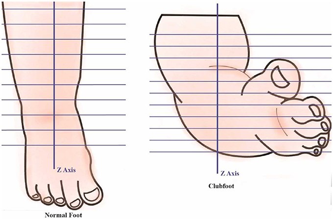
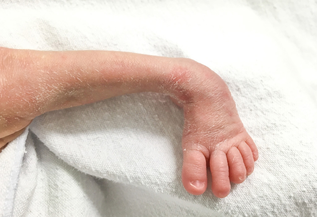
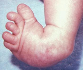
The foot can't be moved into a normal position Clubfoot can affect one or both feet Clubfoot symptoms If your baby has clubfoot, his foot points downwards and inwards like a golf club The middle section of your baby's foot also twists inwards, which makes the foot Club Foot Talipes equinovarus (once called club foot) is a deformity of the foot and ankle that a baby can be born with It is not clear exactly what causes talipes In most cases, it is diagnosed by the typical appearance of a baby's foot after they are born The Ponseti method is now a widely used treatment for talipesThis Pin was discovered by Danielle Brown Discover (and save!) your own Pins on


The Clubfoot Chronicles The Saga Begins


1
Club Foot Symptoms In the majority of cases, the deformity twists the very top of the baby's foot inward and downward, which turns the heel inmost as well as increases the arch This foot can be twisted so seriously that in some cases it appears as if it is upsidedown The foot was in a funny position in A clubfoot would not affect the course of the Horger cited the case of a woman who received an ultrasound diagnosis that her fetus had noUltrasound of the Foot and Ankle



Clubfoot Deformity Symptoms And Treatment Orthoindy Blog


Congenital Talipes Equinovarus Radiology Case Radiopaedia Org
Kawashima T, Uhthoff HK Development of the foot in prenatal life in relation to idiopathic club foot J Pediatr Orthop 1990;The diagnosis was changed after followup ultrasound scan in 13 fetuses (25%), and the final ultrasound diagnosis was normal in one fetus, isolated club foot in 31 fetuses, and complex club footShe's healthy, baby girl is measuring a bit ahead size wise too!
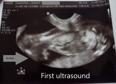


My Journey With Baby S Positional Clubfoot Part 1 Baby Gizmo


Correlations Between Physical And Ultrasound Findings In Congenital Clubfoot At Birth Sciencedirect
Talipes (Club Foot) Club Foot is an abnormality where the feet take on an abnormal position in reference to the lower leg Most commonly the foot is turned inwards and upwards This may affect either one or both feet to varying degrees It is a relatively common fetal malformation and occurs in 1 in 500 to 1 in 1000 live birthsThere are a few birth defects that can be accompanied by clubfeet, so we recommend a thorough exam of your baby using a level II ultrasound Isolated clubfeet will not affect your pregnancy However, if your child has another birth defect that accompanies clubfeet, you may need more frequent monitoring to evaluate your child's wellbeing during the pregnancyUltrasound equipment has a wide array of options and features These features are typically operated from either the console of the ultrasound , a touch screen monitor or a equipment combination of both Figure 2( 9) The basic controls that you need to familiarize yourself in the early stages of ultrasound scanning are the following


A Peachtree City Life An Update On Our Baby Girl Clubfoot


Before Going To Doctor Which Must Know About Clubfoot Rxharun
Figure 4 Radial ray aplasia with club hand in a trisomy 18 fetus at 19 weeks Another finding of trisomy 21 is elevation of the first toe This can be seen by ultrasound because the first toe is no longer in the same plane as the other toes In a sagittal section one can see the angle between the big toe and the rest of the foot Club Foot Reviewed on Twenty though it can sometimes be seen during a fetal ultrasound in the womb While it can be very upsetting to learn that your baby has a deformity, During treatment, an orthopedic surgeon stretches your baby's foot into the correct position and then casts the leg from the footJust had our anatomy scan, I'm w5d Doctor said everything looks good &


Clubfoot In Newborns Paedicare Paediatricians


Prenatal Ultrasound Diagnosis Of Club Foot The Bone Joint Journal
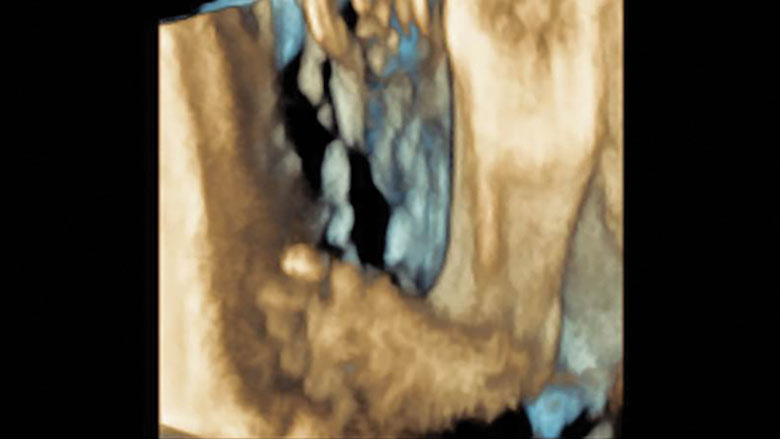
This congenital anomaly is seen in one out of every 1,000 babies, with half of the cases of club foot involving only one foot There is currently no known cause of idiopathic clubfoot, but baby boys are twice as likely to have clubfoot compared to baby girls Neurogenic Clubfoot Neurogenic clubfoot is caused by an underlying neurologic condition3D ultrasound of the same fetus shows the downward pointing toes and lifted heels to better advantage A closeup lateral view of the foot in a fetus with a clubfoot shows marked plantar flexion The image is reminiscent of a ballerina in pointe shoes, which is not a normal foot position for a fetusBasit S, Khoshhal KI Genetics of clubfoot;


About Clubfoot
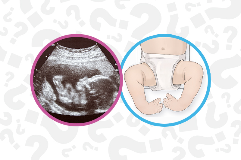


When Your Baby Has Clubfoot Answers For Expecting Parents Boston Children S Answers
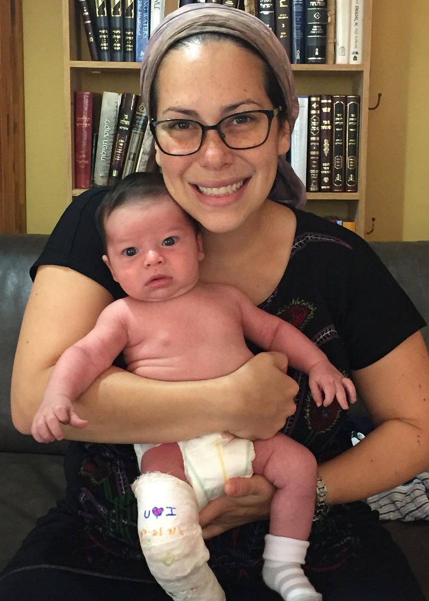
Ftm here and tomorrow I'll be 21 weeks We had our anatomy scan at 19w 3d We got asked to come back in this past Tuesday because doctor wanted to discuss our ultrasound She explained that the tech could not get a good view of baby's feet so they thought there could be a possibility of club footClub foot on ultrasound anyone else?Serial casting The foot is gradually stretched into the correct position and then held in place with casts These casts are changed at the clinic once a week for about 68 weeks Tenotomies (tendon lengthening) In the majority of cases, after we have corrected as much of the foot as possible through casting, your child may require a heel cord (Achilles) tenotomy
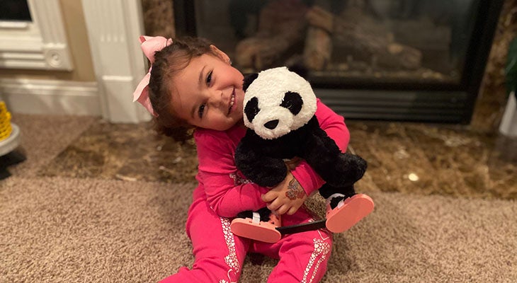


Family Embraces Clubfoot Treatment For Daughter Norton Children S Louisville Ky


1
Perhaps the baby is shy But, honestly, babies really have no concept of being spied on when the ultrasound equipment is turned on It's hard to know the 'why', but some babies have a personal preference for comfort Try not to worry, especially since you have no other markers Most trisomy babies they will also look for club foot, heart conditions, baby size and at this time it's normal for baby to measure low or high I had one at 17 weeks they said baby measured almost two weeks back then one at weeks and baby measured over Club Foot and Pregnancy If club foot is detected during a pregnancy ultrasound, you may want to speak with the pediatrician who will be caring for your infant after birth about the condition Early treatment can prevent the need for surgery Life Expectancy Club foot does not reduce life expectancy


Club Foot


10 Ut Dms Ideas Medical Ultrasound Ultrasound Technician Diagnostic Medical Sonography
Wang JH, Palmer RM, Chung CS The role of major gene in clubfoot Am J Hum Genet 19;Ultrasound of the Ankle and FootIs easily corrected, but hubby &


Congenital Talipes Equinovarus Radiology Reference Article Radiopaedia Org
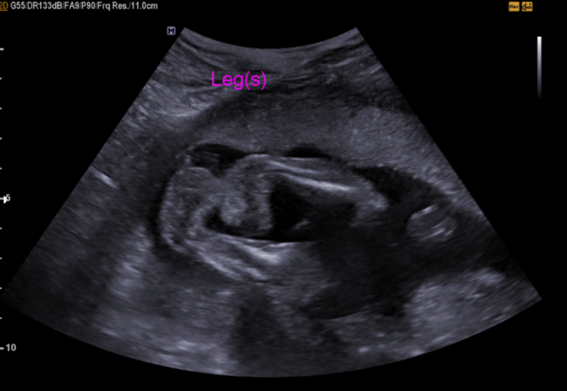


Podopeds Clubfoot Flashcards Memorang
The Fetal Medicine Foundation is aware of the General Data Protection Regulation and changes to data protection legislation This is one of a number of legislative requirements that we must adhere to and as part of the service that you receive from usClubfoot is a congenital foot deformity that affects a child's bones, muscles, tendons, and blood vessels The front half of an affected foot turns inward and the heel points down In severe cases, the foot is turned so far that the bottom faces sideways or up rather than down The condition, also known as talipes equinovarus, is fairly common Free, official coding info for 21 ICD10CM O358XX0 includes detailed rules, notes, synonyms, ICD9CM conversion, index and annotation crosswalks, DRG grouping and more
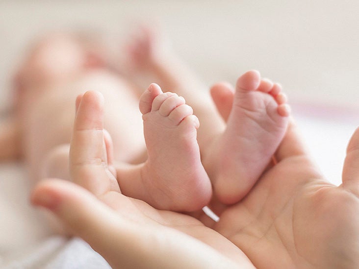


Clubfoot Causes Symptoms And Diagnosis


Ssrd Interesting Cases Fetal Clubfoot Ultrasound Image 3d Image
Anng23 member December 15 in May 16 Moms Had my week anatomy scan a few days ago and they found that the lil one has a mild right club foot All my genetic screens came out in the lowest risk category but I'm still nervous as it's a soft marker for other issuesRecent progress and future perspectives Eur J Med Genet 18;Does anyone know about club feet, so maybe have delt with it before This new ultrasound doctor said that because she couldnt see the foot clearly that she "SUSPECTS" that it MIGHT be a club foot, but also could be the position of the baby, because the baby seems pretty crammed in my small uterous, and i have to go to the hospital for a follow up ultrasound in 3 days?
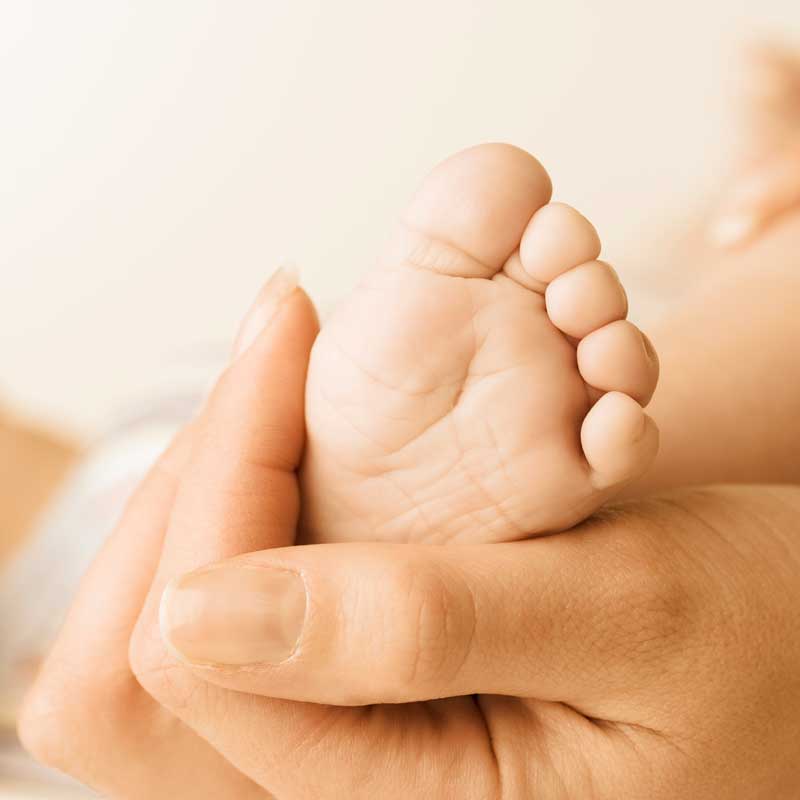


Clubfoot A Mother S Story


The Clubfoot Chronicles The Saga Begins
Clubfoot, or talipes equinovarus, is a deformity in which the foot is excessively plantar flexed, with the forefoot bent medially and the sole facing inwardThis usually results in the underdevelopment of the soft tissues on the medial side of the foot and calf and to various degrees of rigidity of the foot Club foot affects about 1 baby in every 1,000 born in the UK Both feet are affected in about half of these babies It's more common in boys Diagnosing club foot Club foot is usually diagnosed after a baby is born, although it may be spotted during the routine ultrasound scan carried out between 18 and 21 weeks


Club Foot


Ultrasound Video Showing Fetal Anomalies Clubfoot Encephalocele Kyphosis And Placental Mass Youtube
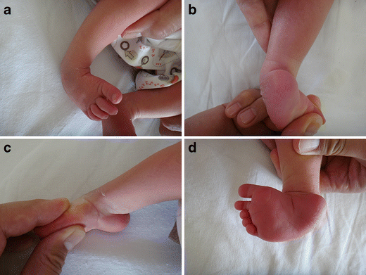


Congenital Clubfoot Early Recognition And Conservative Management For Preventing Late Disabilities Springerlink


First Trimester Physiological Development Of The Fetal Foot Position Using Three Dimensional Ultrasound In Virtual Reality Bogers 19 Journal Of Obstetrics And Gynaecology Research Wiley Online Library
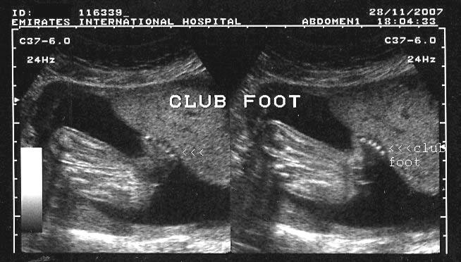


A Gallery Of High Resolution Ultrasound Color Doppler 3d Images Fetal Spine


Clubfoot In Newborns Causes Symptoms Diagnostics Schoen Clinic


Clubfoot Clubfoot Pediatric Interesting Cases And Mcqs Facebook
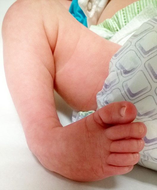


Clubfoot Johns Hopkins Medicine
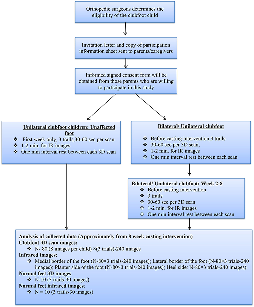


Frontiers Developing A Three Dimensional 3d Assessment Method For Clubfoot A Study Protocol Physiology


Correlations Between Physical And Ultrasound Findings In Congenital Clubfoot At Birth Sciencedirect
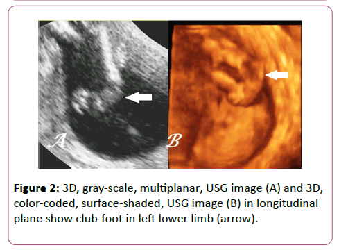


Antenatal 3d Usg In Unilateral Club Foot A Rare Anomaly Insight Medical Publishing


Ssrd Interesting Cases Fetal Clubfoot Ultrasound Image 3d Image
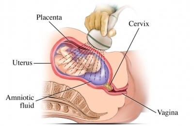


Clubfoot Cook Children S


How Parents And The Internet Transformed Clubfoot Treatment Shots Health News Npr


Three Dimensional 3d Ultrasound Studies A Bilateral Club Foot Download Scientific Diagram


The Ultimate Guide To A Clubfoot Baby Simply Working Mama
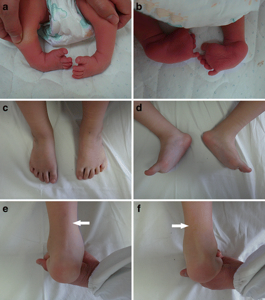


Congenital Clubfoot Early Recognition And Conservative Management For Preventing Late Disabilities Springerlink
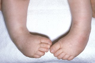


Club Foot Nhs


Clubfoot Versus Positional Foot Deformities On Prenatal Ultrasound Imaging Brasseur Daudruy Journal Of Ultrasound In Medicine Wiley Online Library


Clubfoot In Children Lurie Children S


Frontiers Developing A Three Dimensional 3d Assessment Method For Clubfoot A Study Protocol Physiology


Ponseti Method Archives Steps


Club Foot Talipes Equinovarus Ankle Foot And Orthotic Centre


Just Had Anatomy Scan What Do Y All Think Clubfoot Forums What To Expect


404 Not Found Ultrasound Sonography Ultrasound Sonography


Clubfoot Deformity Talipes Equinovarus


Club Foot


Club Foot In Infants Reasons Signs Remedies


Club Foot Antenatal Ultrasound Radiology Case Radiopaedia Org


A Step In The Right Direction Treating Clubfoot Sans Surgery Health Beat Spectrum Health


Club Foot Nhs


Skeleton Diagnosis Of Fetal Abnormalities The 18 23 Weeks Scan


Diagnosis Of Fetal Syndromes By Three And Four Dimensional Ultrasound Is There Any Improvement


The Clubfoot Chronicles The Saga Begins


Clubfoot Causes And Treatments


Clubfoot Paley Orthopedic Spine Institute


Clubfoot Symptoms And Causes Mayo Clinic


Clubfoot Boston Children S Hospital


Clubfoot Talipes Equinovarus Radiology Key


June 3rd World Clubfoot Day


Pdf Prenatal Ultrasound Diagnosis Of Club Foot Outcome And Recommendations For Counselling And Follow Up Semantic Scholar


Clubfoot Treatment At Kids Orthopedic Kolkata By Kids Orthopedic Issuu


Ultrasound Video Showing Club Foot Fetal Anomaly Scan Youtube


Congenital Talipes Equinovarus Radiology Reference Article Radiopaedia Org


Club Foot Talipes In Babies Causes Signs Treatment Youtube


Clubfoot Orthopaedia


Clubfoot Deformity Talipes Equinovarus


Clubfoot Versus Positional Foot Deformities On Prenatal Ultrasound Imaging Brasseur Daudruy Journal Of Ultrasound In Medicine Wiley Online Library


Clubfoot Healthdirect


Pdf Prenatal Ultrasound Diagnosis Of Club Foot Outcome And Recommendations For Counselling And Follow Up Semantic Scholar


Club Foot


Clubfoot Curved Baby Feet Next Step Foot Ankle Clinic


Overcoming Clubfoot One Mom S Story Parents


Prenatal Ultrasound Diagnosis Of Club Foot The Bone Joint Journal


My Journey With Baby S Positional Clubfoot Part 1 Baby Gizmo


Club Foot In Ultrasound Babycenter


Tackling Talipes Early With A Team Approach Children S Hospital Of Philadelphia


Clubfeet At 12 Weeks A Transabdominal Scan At 12 Weeks 4 Days Shows Download Scientific Diagram


The Ultrasound Images Of Fetus Diagnosed As Clubfoot Download Scientific Diagram


Clubfoot Deformity Talipes Equinovarus


Overcoming Clubfoot One Mom S Story Parents


Talipes Equinovarus Club Ultrasound Guided Tips Facebook


Clubfoot Deformity Talipes Equinovarus


1


Pdf Congenital Talipes Equinovarus A Case Report Of Bilateral Clubfoot In Three Homozygous Preterm Infants Semantic Scholar
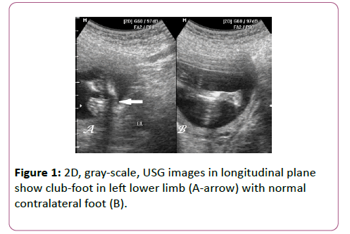


Antenatal 3d Usg In Unilateral Club Foot A Rare Anomaly Insight Medical Publishing


Kentucky Family Describes Clubfoot Treatment Process Whas11 Com


Club Foot Pediatric Ortho
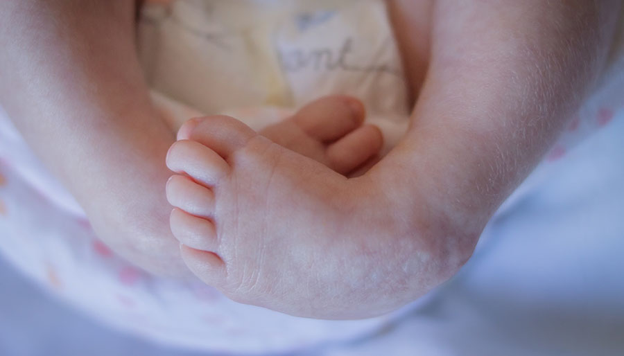


Marlowe S Clubfoot Journey How One Mom Went From Devastated To Reassured Children S Wisconsin
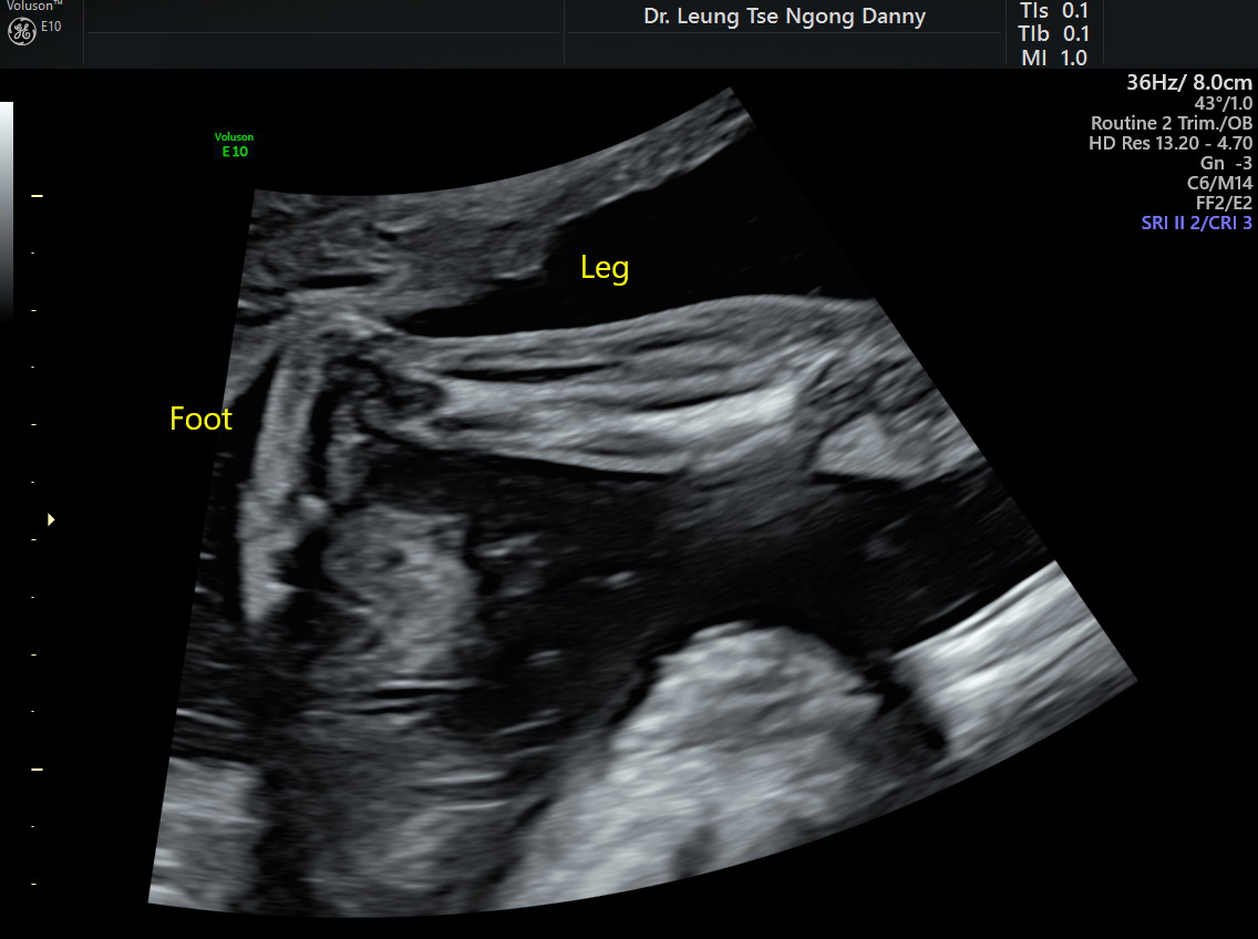


Club Foot Congenital Talipes Equinovarus Hkog Info


Clubfoot At 22 Second Weeks Gestation The Forefoot Arrows Is Download Scientific Diagram


Clubfoot Boston Children S Hospital


Fetus General Normal Fetal Anatomy Ultrasound Services Service Provider From Ernakulam


How Parents And The Internet Transformed Clubfoot Treatment Shots Health News Npr


Predicting Recurrence After Clubfoot Treatment Lower Extremity Review Magazine


Clubfoot Versus Positional Foot Deformities On Prenatal Ultrasound Imaging Brasseur Daudruy Journal Of Ultrasound In Medicine Wiley Online Library


Clubfoot Causes And Treatments Palos Hills And Mokena


Protected Blog Log In Club Foot How To Apologize Ultrasound


0 件のコメント:
コメントを投稿